Home

kotiček kazalo Napihne fluorescent microscopy markers Frank Worthley Urnik Oče

Fluorescence imaging of cell surface markers. HBMSCs were... | Download  Scientific Diagram
Fluorescence imaging of cell surface markers. HBMSCs were... | Download Scientific Diagram

Precise, Correlated Fluorescence Microscopy and Electron Tomography of  Lowicryl Sections Using Fluorescent Fiducial Markers - ScienceDirect
Precise, Correlated Fluorescence Microscopy and Electron Tomography of Lowicryl Sections Using Fluorescent Fiducial Markers - ScienceDirect

General 2 — Jackson Lab

Detection of fluorescent markers by confocal laser scanning microscopy... |  Download Scientific Diagram
Detection of fluorescent markers by confocal laser scanning microscopy... | Download Scientific Diagram
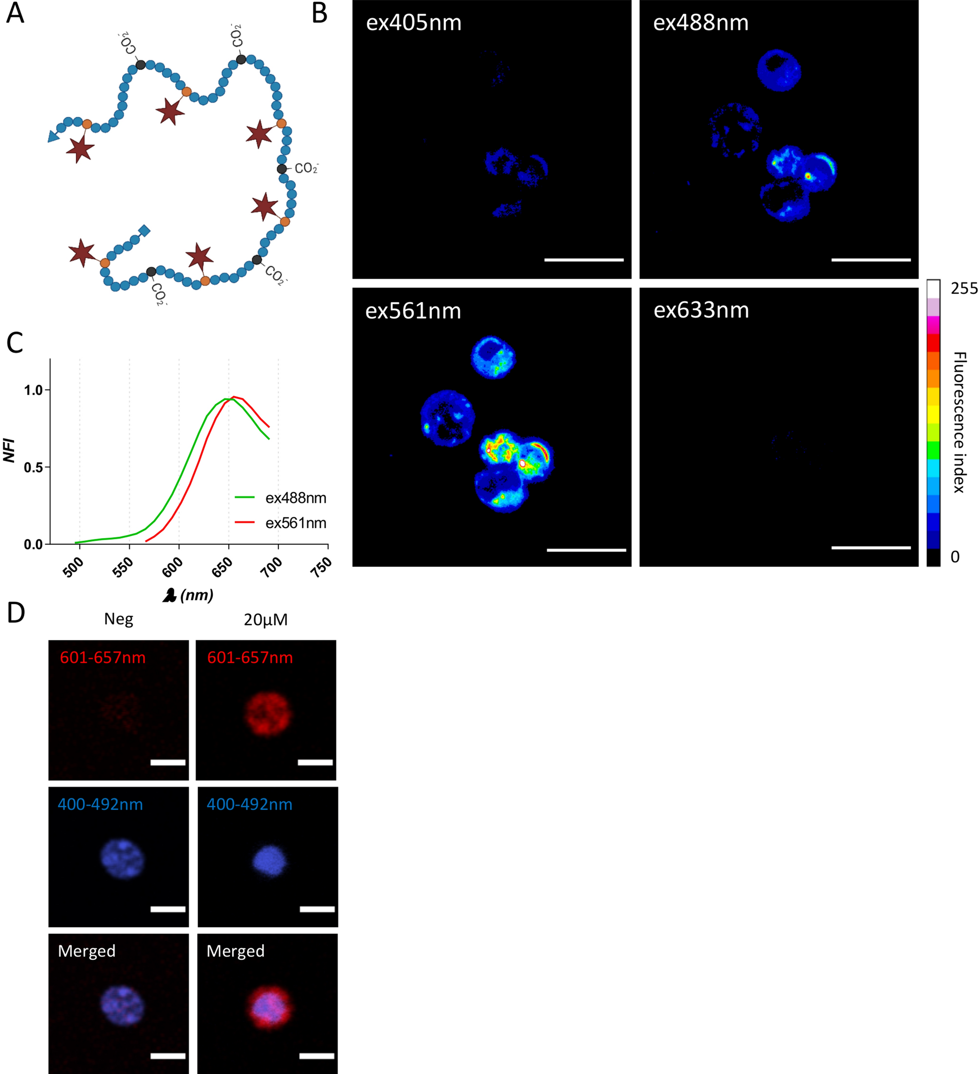
Fluorescence microscopy analysis for expression of cell markers of... |  Download Scientific Diagram
Fluorescence microscopy analysis for expression of cell markers of... | Download Scientific Diagram

Multiscale fluorescent tracking of immune cells in the liver with a highly  biocompatible far-red emitting polymer probe | Scientific Reports
Multiscale fluorescent tracking of immune cells in the liver with a highly biocompatible far-red emitting polymer probe | Scientific Reports

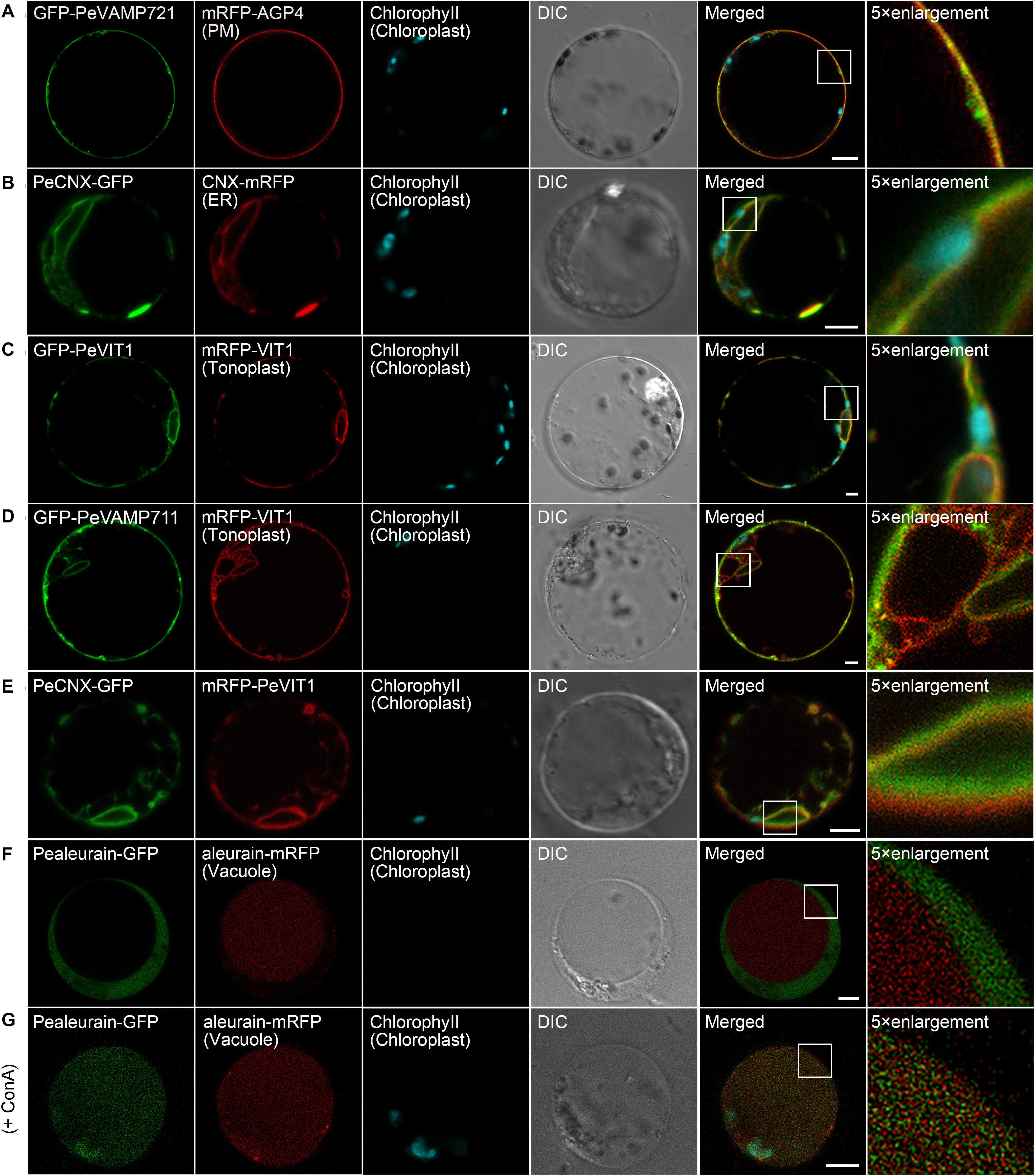
Frontiers | Organelle Visualization With Multicolored Fluorescent Markers  in Bamboo
Frontiers | Organelle Visualization With Multicolored Fluorescent Markers in Bamboo

Introduction: Fluorescence Microscopy - Soft Matter Physics Division -  University of Leipzig
Introduction: Fluorescence Microscopy - Soft Matter Physics Division - University of Leipzig

Fluorescent Dyes | Science Lab | Leica Microsystems

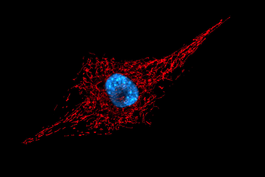
Live mitochondria seen in unprecedented detail: photobleaching in STED  microscopy overcome
Live mitochondria seen in unprecedented detail: photobleaching in STED microscopy overcome

Colocalization of fluorescent markers in confocal microscope images of  plant cells | Nature Protocols
Colocalization of fluorescent markers in confocal microscope images of plant cells | Nature Protocols
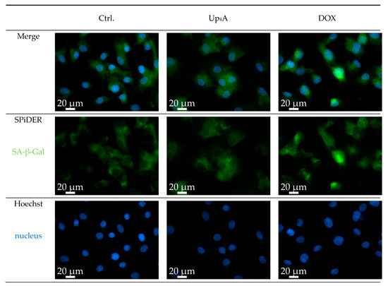
IJMS | Free Full-Text | A Novel Protocol for Detection of Senescence and  Calcification Markers by Fluorescence Microscopy
IJMS | Free Full-Text | A Novel Protocol for Detection of Senescence and Calcification Markers by Fluorescence Microscopy
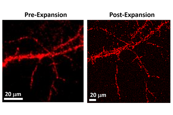
Solved Fluorescence microscopy using antibodies tagged with | Chegg.com

Brighter fluorescent markers allow for finer imaging of nanoscopic objects  | McKelvey School of Engineering at Washington University in St. Louis
Brighter fluorescent markers allow for finer imaging of nanoscopic objects | McKelvey School of Engineering at Washington University in St. Louis

Fluorescent Dyes | Science Lab | Leica Microsystems

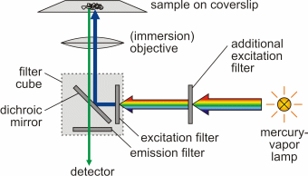
2.3. Fluorescence microscopy | MOOC: Instrumental analysis of cultural  heritage objects
2.3. Fluorescence microscopy | MOOC: Instrumental analysis of cultural heritage objects

Confocal Microscopy - The Fluorescent Protein Color Palette | Olympus LS

A Validated Set of Ascorbate Peroxidase-Based Organelle Markers for  Electron Microscopy of Saccharomyces cerevisiae | mSphere
A Validated Set of Ascorbate Peroxidase-Based Organelle Markers for Electron Microscopy of Saccharomyces cerevisiae | mSphere

Introduction to the Quantitative Analysis of Two-Dimensional Fluorescence  Microscopy Images for Cell-Based Screening | PLOS Computational Biology
Introduction to the Quantitative Analysis of Two-Dimensional Fluorescence Microscopy Images for Cell-Based Screening | PLOS Computational Biology

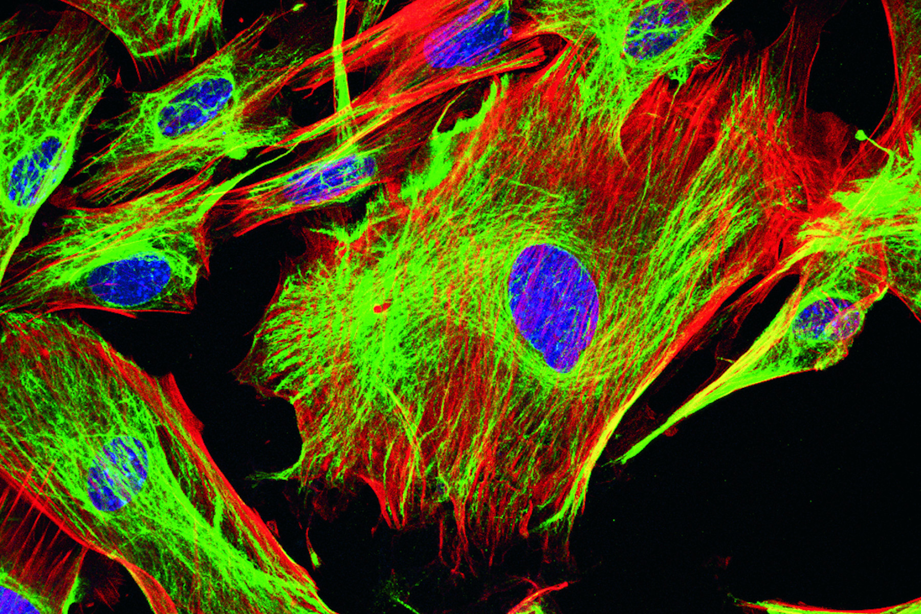
Fluorescent image of human stem cells stained with monoclonal antibodies  markers under the microscopy showing nuclei in blue and microtubules in red  Stock Photo - Alamy
Fluorescent image of human stem cells stained with monoclonal antibodies markers under the microscopy showing nuclei in blue and microtubules in red Stock Photo - Alamy

The Fluorescence Microscope - Preparation of Specimen

Ecole des Houches: Fluorescence markers for advanced microscopy -  France-BioImaging
Ecole des Houches: Fluorescence markers for advanced microscopy - France-BioImaging

Improved Plasmids for Fluorescent Protein Tagging of Microtubules in  Saccharomyces cerevisiae - Markus - 2015 - Traffic - Wiley Online Library
Improved Plasmids for Fluorescent Protein Tagging of Microtubules in Saccharomyces cerevisiae - Markus - 2015 - Traffic - Wiley Online Library